Whitefish Blastula Slide. Solution for biology 12th edition chapter 8, problem 18. About press copyright contact us creators advertise developers terms privacy policy & safety how youtube works test new features press copyright contact us creators.
Tour of the Whitefish Blastula Slide YouTube from www.youtube.com
Look at mitosis models 2. Solution for biology 12th edition chapter 8, problem 18. Notice that the section is a circle composed of dozens of closely packed individual cells.
You Should See Animal Cells In Various Stages Of Mitosis.
This cell is in very late stage of metaphase. Notice that the section is a circle composed of dozens of closely packed individual cells. About press copyright contact us creators advertise developers terms privacy policy & safety how youtube works test new features press copyright contact us creators.
Calculate Percentage Of Each Phase
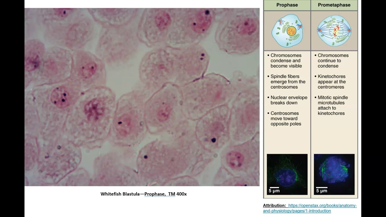
The whitefish slide is from a blastula, an early stage in embryonic development. Whitefish blastula the cells of a creating embryo are dividing quickly and can be utilized for viewing the totally different phases of mitosis. At 1000x, micrograph width = 0.08 mm.
Focus On One That Is In Good Condition And In Which The Cells Are Easy To See.
Put in onion root tip slide. This cell is in the interphase stage of the cell cycle. At 100x, micrograph width = 0.8 mm;
The Whitefish Cells Pictured Below Are Blastula Cells.
Repeat steps 2 and 3 using whitefish blastula slide. Observe whitefish blastula cells under microscope (animal cell mitosis) 3. Interphase, prophase, metaphase, anaphase, and telophase.
Whitefish Blastula The Cells Of A Developing Embryo Are Dividing Rapidly And Can Be Used For Viewing The Different Stages Of Mitosis.
The cells of a blastula, or developing embryo, divide rapidly, which make this whitefish slide useful for viewing the different stages of mitosis. Interphase, prophase, metaphase, anaphase, and telophase. Leave a reply cancel reply.